A common question in dentistry is simple but important: what does a wisdom tooth coming in look like when it is erupting normally, and when does that appearance suggest a problem? Wisdom teeth, also called third molars, often come in later than other teeth and may have less room to erupt.
In many people, a wisdom tooth coming in looks like a small white cusp breaking through the gum at the very back of the mouth. Sometimes only one corner is visible, while the rest of the tooth stays covered by gum tissue.
Other times, you may not see much tooth at first. The gum behind the last molar may look raised, stretched, slightly swollen, or lighter in color where the tooth is pushing upward.
Early eruptions can be subtle. You might notice a new ridge under the gum, mild tenderness when brushing, or a flap of tissue over a partially erupted tooth.
South Florida Sedation Dentistry offers comprehensive dental exams in Greenacres, FL, and can evaluate erupting wisdom teeth.
Wisdom teeth are the last permanent teeth to erupt, usually in the late teens or twenties. By that point, the jaw has already made room for the rest of the adult teeth, so third molars may have limited space.
That is why a wisdom tooth may come in looking uneven, partly hidden, or tilted toward the tooth next to it. This does not always mean something is wrong, but it does mean the area should be watched closely.
A fully erupting wisdom tooth may look like any other molar, with several rounded chewing points visible above the gumline. A partially erupting wisdom tooth often looks less defined because only the top edge or one cusp can be seen.
If a wisdom tooth is coming in, the most noticeable change is usually behind the last regular molar. One side may change before the other, since eruption is not always symmetrical.
You may notice:
Not every erupting wisdom tooth is easy to see. Some stay fully under the gum or bone, which is called impaction when eruption is blocked or incomplete.
A tooth may also erupt at an angle. In that case, the visible part may seem to lean forward or come in sideways instead of rising straight up.
The most common problem around a partially erupted wisdom tooth is inflammation of the gum tissue over it. Dentists call this pericoronitis, which means inflammation around the crown, or visible top, of a tooth.
The gum may look more than mildly swollen. It can appear red, shiny, tender, and thickened, and a flap of tissue may trap plaque, food debris, and bacteria. If left untreated, this can increase the risk of gum disease.
In some cases, the area develops painful swelling around a partially erupted wisdom tooth instead of a quiet, gradual eruption. That is more concerning when there is a bad taste, bad breath, pus, difficulty chewing, or pain when opening the mouth.
These findings do not always mean a serious infection, but they do justify prompt dental evaluation. The back of the mouth is hard to inspect and clean well, so irritation can worsen faster than many people expect.
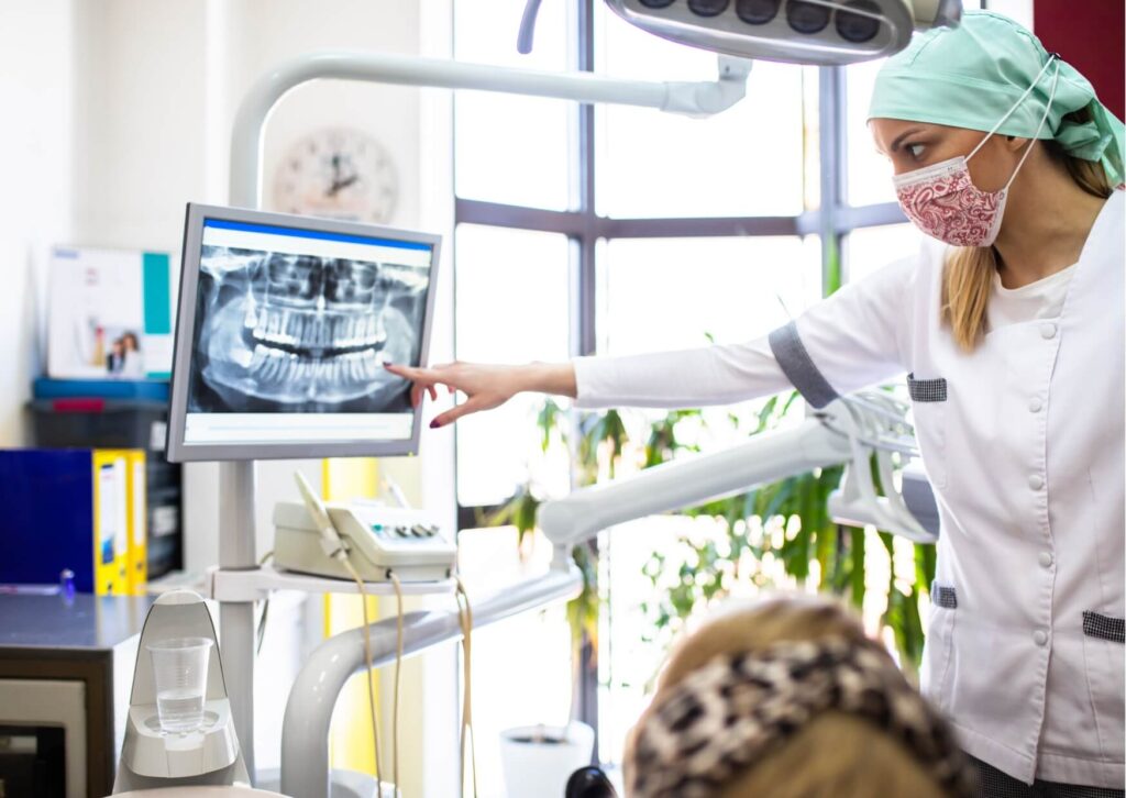
What you see in the mirror does not always match what is happening below the surface. A wisdom tooth may look like it is barely coming in, while an X-ray shows that much more of the crown has formed and may be pressing against the tooth in front of it.
Wisdom teeth can also change position over time. Some gradually move into a better position, while others stay impacted in bone or soft tissue.
That is why a visual check alone has limits. If the area is painful, repeatedly inflamed, hard to clean, or unclear on exam, a dental exam and X-ray can help determine whether the tooth is erupting normally, partially impacted, or affecting the neighboring molar.
A wisdom tooth may be erupting in a fairly normal way if the changes are mild, gradual, and not linked to significant pain or repeated swelling. The gum may feel tender for a short time, and a small part of the tooth may become visible over weeks or months.
Normal does not always mean comfortable. It means the tooth appears to be progressing without signs of infection, damage to nearby teeth, or ongoing tissue trapping.
Typical features of a more routine eruption include:
Even with milder findings, regular dental follow-up is a good idea. Wisdom teeth can change position over time, and a tooth that seems manageable now may become harder to clean later.
Some changes suggest the issue is no longer a simple eruption. A dentist should assess the area promptly if there is worsening pain, significant swelling, repeated gum infection, or concern that the tooth is pushing into the molar in front of it.
Seek prompt dental care or emergency care if you notice:
If swelling affects breathing, swallowing, or your ability to open the mouth normally, urgent in-person care is important. Those symptoms can point to a spreading infection and should not be monitored at home.
Treatment depends on what the tooth looks like on exam and on imaging. In many cases, the options are monitoring, improving access for cleaning, or planning removal if the tooth is impacted, repeatedly inflamed, or threatening the adjacent molar.
Appearance alone does not decide treatment. The pattern over time matters more, including repeated inflammation, trapped debris, decay risk, gum disease risk, and the position of the tooth.
Sometimes a partly visible wisdom tooth can stay stable for years. In other cases, a tooth that shows only a small white edge is already creating a long-term hygiene problem and is unlikely to erupt into a useful position.
When removal or more complex management is needed, referral for oral surgery is common. For patients who feel nervous about treatment, sedation options can make care more comfortable.
The ADA also publishes sedation guidelines for dentists. How sedation works is explained in our guide: How sedation works. IV sedation is described in more detail in a dedicated overview: IV sedation.
A careful dental exam is more useful than trying to judge the area from a mirror alone. The visible changes may start the question, but the full answer usually comes from the exam, the X-ray, and how the area behaves over time.
If you are noticing a new tooth edge, swelling, or a gum flap at the back of the mouth, it is reasonable to have the area checked before irritation becomes a larger problem.
South Florida Sedation Dentistry in Greenacres, FL offers dental exams and can help patients from Wellington and Royal Palm Beach. Call us at (561) 967-2001 to schedule.
At first, it may look like a small white point or ridge under or through the gum behind the last molar. Sometimes the first visible change is not the tooth itself but a swollen or raised patch of gum.
Yes. A wisdom tooth may be moving upward under the gum with only subtle swelling or tenderness visible. In some cases, very little can be seen without an X-ray.
It can happen during a partial eruption and is fairly common. However, that flap can trap bacteria and food, so it should be evaluated if it becomes painful, swollen, or repeatedly irritated.
Not always, but a sideways or tilted eruption pattern raises concern for impaction and cleaning difficulty. A dentist usually needs an exam and X-ray to decide whether monitoring or removal makes more sense.
You should seek dental care if there is increasing pain, spreading swelling, drainage, fever, trouble opening the mouth, or repeated infection around the area. Those signs suggest more than a routine eruption.
Have your friend, family member, or co-worker scan the code below or visit southfloridasedationdentistry.com on their phone to load our site. Most modern cell phones can scan QR codes using the phone’s camera.